Précis
Aucun mouvement
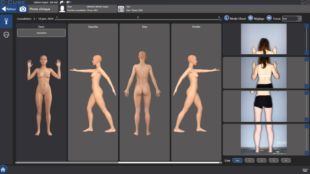
Repositionnement parfait du corps entier

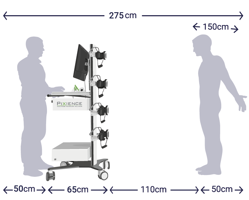
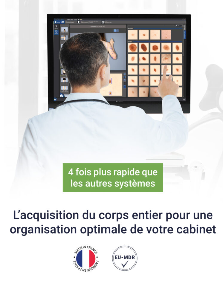
Rapide
4 fois plus rapides que tous les autres systèmes grâce aux 4 caméras fixes

Confortable
Réduction du temps de maintient de posture du patient

Silencieux
Système ultra-silencieux grâce à l'absence de motorisation
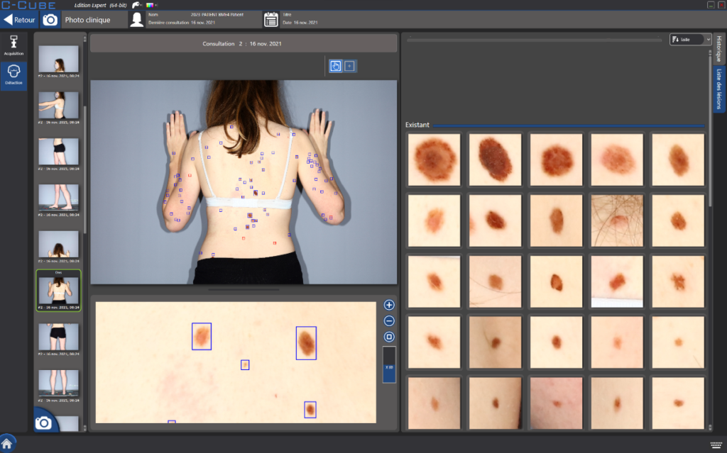
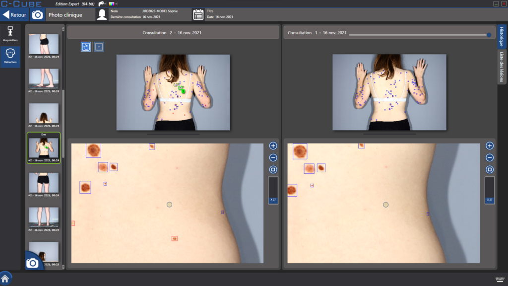
Précis
Aucun mouvement
Repositionnement parfait du corps entier
Confortable
Réduction du temps de maintient de posture du patient
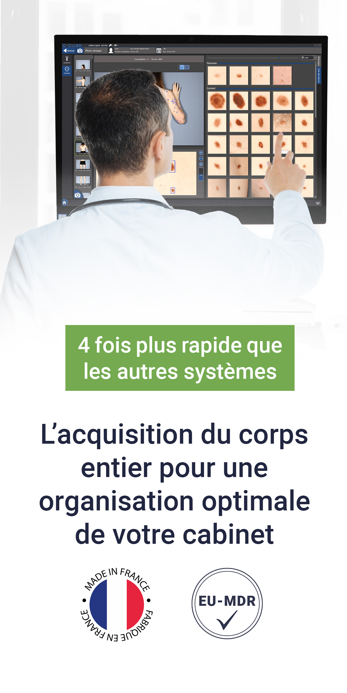
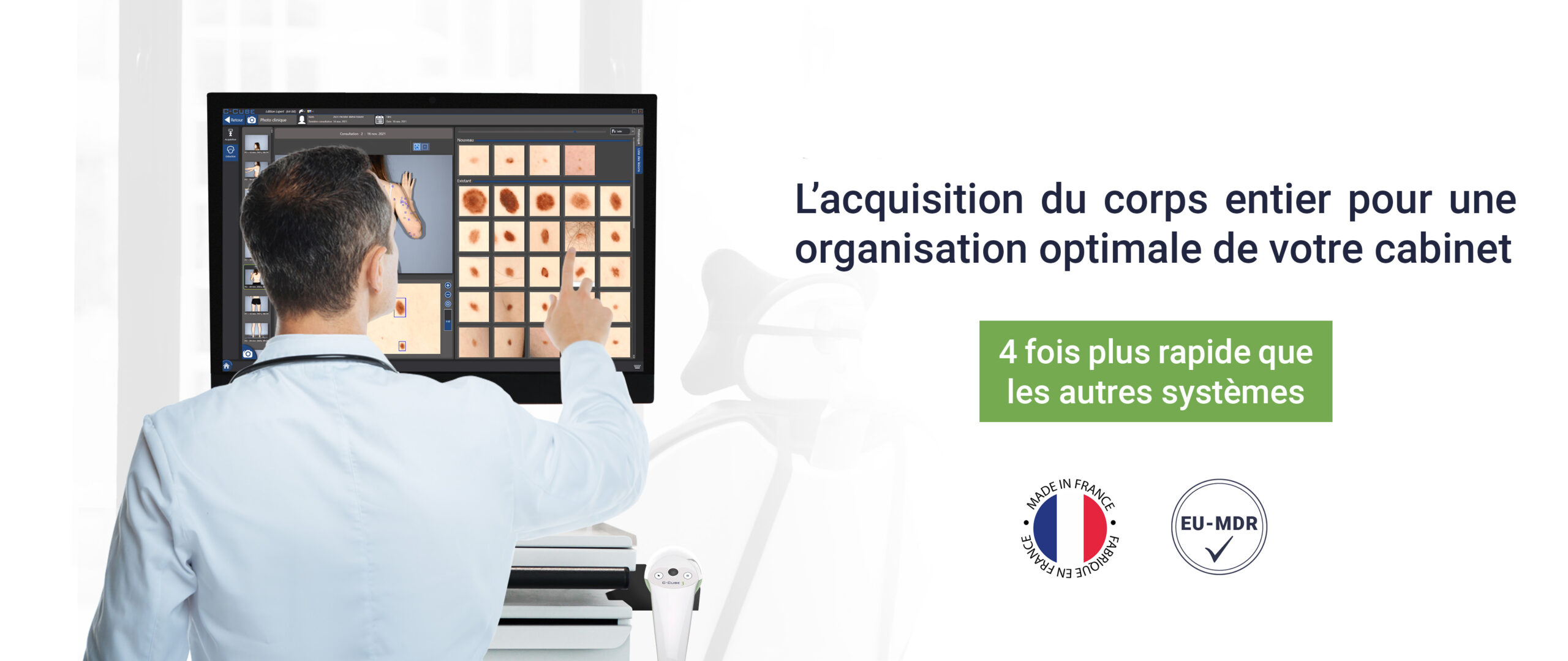
Rapide
4 fois plus rapides que tous les autres systèmes grâce aux 4 caméras fixes
Silencieux
Système ultra-silencieux grâce à l'absence de motorisation